When a doctor says you need a scan, it often comes with a flurry of unfamiliar terms — MRI, CT, ultrasound, contrast, fasting, radiation. For most people, the instinct is to nod along and worry later. But understanding what each type of imaging actually does, why one might be chosen over another, and how to prepare for it can turn an anxious experience into a straightforward one. Here’s a clear, jargon-free guide to the four imaging tools you’re most likely to encounter.
Why there are different types of scans
No single imaging technology can see everything. Each one works on different physics and is suited to different parts of the body. Bone, soft tissue, blood flow and moving organs all “show up” differently depending on the method used. That’s why a doctor’s choice of scan is deliberate — they’re matching the tool to the question they’re trying to answer. Knowing the basics helps you understand your own care rather than feeling like a passenger in it.
X-ray: fast, simple and everywhere
The X-ray is the oldest and most widely used imaging method, and for good reason. It uses a small, controlled dose of radiation to produce a flat image, and it excels at showing dense structures — particularly bone. If you’ve fractured a wrist, twisted an ankle badly, or have a suspected chest infection, an X-ray is usually the first step.
Its strengths are speed and accessibility: the image takes only seconds to capture, and there’s rarely any preparation involved. Its limitation is detail — X-rays are excellent for bone and certain chest conditions, but they don’t reveal soft tissues like muscles, ligaments or internal organs in any depth. When a doctor needs that level of detail, they move to a more advanced method.
CT scan: cross-sectional clarity, fast
A CT (computed tomography) scan is essentially a sophisticated evolution of the X-ray. Instead of one flat image, it takes many X-ray slices from different angles and uses a computer to combine them into detailed cross-sectional views of the body. The result is far richer information about bones, blood vessels and soft tissues alike.
CT is prized for its speed and is often the imaging of choice in emergencies — trauma, sudden abdominal pain, suspected internal bleeding or stroke assessment — because it can scan large areas of the body in under a minute. Sometimes a contrast dye is used to make blood vessels or specific tissues stand out more clearly. Because CT uses X-ray radiation, doctors weigh its use thoughtfully, but modern machines use low-dose techniques, and in the right situation the diagnostic benefit far outweighs the small risk.
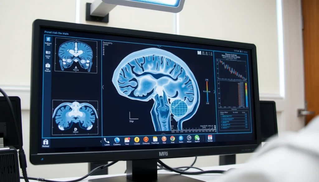
MRI: unmatched soft-tissue detail, no radiation
Magnetic resonance imaging (MRI) takes a completely different approach. Instead of radiation, it uses powerful magnets and radio waves to generate extraordinarily detailed images — particularly of soft tissues that X-ray and even CT struggle to capture clearly.
This makes MRI the gold standard for the brain and spinal cord, joints and ligaments, muscles, and many conditions affecting internal organs. If you have persistent knee pain, unexplained neurological symptoms, or a suspected disc problem, an MRI scan often provides the clearest answer. Because it uses no ionising radiation, it’s also well suited to situations requiring repeated imaging over time.
The trade-offs are that an MRI takes longer — typically 20 to 45 minutes — and requires you to lie still inside a tunnel-like machine, which can feel confining for those prone to claustrophobia. The strong magnet also means anyone with certain metal implants or devices must be carefully screened beforehand. None of this is cause for alarm; it simply means being upfront about your medical history when you book.
Ultrasound: real-time, completely safe
Ultrasound uses high-frequency sound waves rather than radiation to create live, moving images of what’s happening inside the body. A handheld probe glides over the skin with a layer of gel, and the reflections are translated into a real-time picture.
Its biggest advantages are safety and immediacy. With no radiation involved, ultrasound is the imaging of choice during pregnancy and is widely used to examine the abdomen, kidneys, gallbladder, thyroid and blood flow through vessels. Because it shows movement, it’s also excellent for assessing things that change moment to moment — such as a beating heart or blood circulation. It’s painless, generally requires little preparation, and results are often available very quickly.
How to prepare — and why it matters
Preparation varies by scan, and following the instructions genuinely affects accuracy:
· Fasting is often required for certain abdominal scans and contrast studies — usually a few hours without food.
· A full bladder may be requested for some pelvic and obstetric ultrasounds.
· Metal and devices must be declared before an MRI, including implants, pacemakers and even certain tattoos or makeup containing metallic particles.
· Allergies and kidney health matter when contrast dye is involved, so mention any history of reactions or kidney issues.
When in doubt, ask the facility directly. A two-minute clarifying call beats arriving unprepared and needing to reschedule.
What the results mean
A scan produces images, but interpretation is a specialist skill. Trained radiologists examine the pictures and prepare a report for your treating doctor, who then explains the findings in the context of your symptoms and history. It’s worth remembering that an “incidental finding” — something noticed that’s unrelated to your original concern — is common and often harmless. The right response to any result is a conversation with your doctor, not a deep dive into worst-case scenarios online.
The bottom line
Each imaging method is a different lens on the body: X-ray for quick bone assessment, CT for fast and detailed cross-sections, MRI for unmatched soft-tissue clarity, and ultrasound for safe, real-time views. You don’t need to become an expert — but understanding the basics helps you ask better questions, prepare properly, and approach your scan with confidence rather than apprehension. Good diagnosis begins with the right picture, and now you know how those pictures are made.
